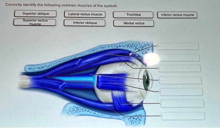
Correctly identify the following extrinsic muscles of the eyeball. – Correctly identifying the extrinsic muscles of the eyeball is essential for understanding eye movement, ophthalmic examinations, and surgical procedures. These muscles, responsible for controlling the direction and coordination of eye movements, play a crucial role in our ability to see and interact with the world around us.
This comprehensive guide will delve into the anatomy, functions, and clinical significance of these muscles, providing a thorough understanding for healthcare professionals and individuals alike.
Extrinsic Muscles of the Eyeball

Extrinsic muscles of the eyeball are responsible for controlling eye movements. These muscles originate from the bony orbit and insert onto the surface of the eyeball. They are innervated by cranial nerves III, IV, and VI.
Muscles Responsible for Eye Movement
- Superior rectus:Elevates the eye
- Inferior rectus:Depresses the eye
- Medial rectus:Adducts the eye
- Lateral rectus:Abducts the eye
- Superior oblique:Intorts and depresses the eye
- Inferior oblique:Extorts and elevates the eye
Location and Attachments
The extrinsic eye muscles are located within the orbit. They originate from the bony orbit and insert onto the surface of the eyeball.
| Muscle | Origin | Insertion |
|---|---|---|
| Superior rectus | Lesser wing of the sphenoid bone | Superior aspect of the eyeball |
| Inferior rectus | Inferior orbital fissure | Inferior aspect of the eyeball |
| Medial rectus | Medial wall of the orbit | Medial aspect of the eyeball |
| Lateral rectus | Lateral wall of the orbit | Lateral aspect of the eyeball |
| Superior oblique | Medial wall of the orbit | Trochlea, then superior aspect of the eyeball |
| Inferior oblique | Inferior orbital fissure | Inferior aspect of the eyeball |
Clinical Significance
A thorough understanding of the extrinsic muscles of the eyeball is essential for ophthalmic examinations, diagnoses, and surgical procedures.
- Ophthalmic examinations:The extrinsic eye muscles are assessed during eye examinations to evaluate their function and identify any abnormalities.
- Diagnoses:Weakness or paralysis of the extrinsic eye muscles can indicate underlying neurological conditions, such as myasthenia gravis or oculomotor nerve palsy.
- Surgical procedures:The extrinsic eye muscles may be surgically manipulated to correct strabismus (misalignment of the eyes).
Functional Anatomy
The extrinsic eye muscles work together to coordinate eye movements, including binocular vision and accommodation.
Binocular vision:The extrinsic eye muscles allow for the precise alignment of both eyes, enabling binocular vision and depth perception.
Accommodation:The extrinsic eye muscles also play a role in accommodation, the ability to focus on objects at different distances.
Comparison with Intrinsic Muscles, Correctly identify the following extrinsic muscles of the eyeball.
| Characteristic | Extrinsic Muscles | Intrinsic Muscles |
|---|---|---|
| Function | Control eye movements | Control pupil size and lens shape |
| Location | Outside the eyeball | Within the eyeball |
| Innervation | Cranial nerves III, IV, and VI | Parasympathetic and sympathetic nerves |
Question & Answer Hub: Correctly Identify The Following Extrinsic Muscles Of The Eyeball.
What are the key differences between extrinsic and intrinsic eye muscles?
Extrinsic muscles are responsible for controlling eye movements, while intrinsic muscles control the pupil size and lens shape.
How do the extrinsic eye muscles work together to achieve coordinated eye movements?
The extrinsic eye muscles work in pairs, with one muscle responsible for moving the eye in one direction and its counterpart responsible for moving it in the opposite direction. This coordinated action allows for precise and efficient eye movements.
What are the potential clinical implications of misidentifying the extrinsic eye muscles?
Misidentifying the extrinsic eye muscles can lead to incorrect diagnoses and inappropriate treatment, potentially affecting visual function and patient outcomes.

