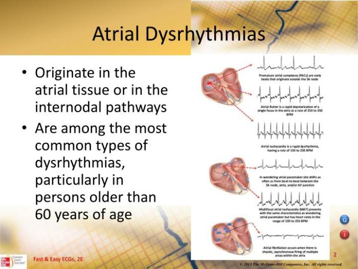
An atrial dysrhythmia originates from the heart’s atria, triggering abnormal electrical impulses that disrupt the regular rhythm of the heart. This comprehensive overview delves into the anatomical structures, electrophysiological mechanisms, clinical manifestations, diagnostic evaluation, treatment options, and prevention strategies associated with atrial dysrhythmias, providing a thorough understanding of this prevalent cardiac condition.
Atrial dysrhythmias arise from various anatomical sites within the atria, including the sinoatrial node, atrioventricular node, and atrial myocardium. The electrophysiological characteristics of each origination site determine the specific type of dysrhythmia, such as atrial fibrillation, atrial flutter, or paroxysmal supraventricular tachycardia.
Atrial Dysrhythmia Origination Sites

Atrial dysrhythmias can originate from various anatomical structures within the atria, including the sinoatrial node (SAN), atrioventricular node (AVN), atrial appendages, Bachmann’s bundle, and coronary sinus.
The SAN, located in the right atrium, is the primary pacemaker of the heart and is responsible for initiating electrical impulses that trigger atrial contractions. Dysrhythmias originating from the SAN are characterized by abnormal heart rates and rhythms, such as sinus tachycardia, sinus bradycardia, and sick sinus syndrome.
Atrioventricular Node (AVN), An atrial dysrhythmia originates from
The AVN, situated between the atria and ventricles, plays a crucial role in delaying electrical impulses before they reach the ventricles. Dysrhythmias originating from the AVN can result in atrioventricular blocks, where the electrical impulses are either delayed or blocked, leading to abnormal heart rhythms and conduction disturbances.
Atrial Appendages
The atrial appendages are small, ear-shaped structures located at the superior aspect of each atrium. Dysrhythmias originating from the atrial appendages are often characterized by atrial fibrillation, a common type of arrhythmia characterized by rapid, irregular atrial contractions.
Bachmann’s Bundle
Bachmann’s bundle is a group of specialized cardiac fibers that connect the right and left atria. Dysrhythmias originating from Bachmann’s bundle can lead to interatrial conduction disturbances, affecting the coordination of electrical impulses between the atria.
Coronary Sinus
The coronary sinus is a large vein that collects blood from the heart. Dysrhythmias originating from the coronary sinus are less common but can include atrial tachycardia, characterized by rapid, regular atrial contractions.
Electrophysiological Mechanisms: An Atrial Dysrhythmia Originates From

Atrial dysrhythmias arise from abnormal electrical impulses within the atria. These impulses can originate from various sites within the atria, leading to different types of dysrhythmias.
The abnormal electrical impulses underlying atrial dysrhythmias can be classified into three main mechanisms: automaticity, triggered activity, and re-entry.
Automaticity
Automaticity refers to the ability of cardiac cells to generate electrical impulses spontaneously. In normal conditions, the sinoatrial node (SA node) is the dominant pacemaker of the heart, generating regular electrical impulses that initiate each heartbeat.
In atrial dysrhythmias, other cells within the atria may develop automaticity, leading to the generation of abnormal electrical impulses. These impulses can compete with the normal impulses from the SA node, causing irregular heartbeats.
Triggered Activity
Triggered activity occurs when an electrical impulse is triggered by an abnormal electrical stimulus, such as a premature depolarization or an early afterdepolarization.
Premature depolarizations are electrical impulses that occur before the expected time. They can be caused by various factors, such as electrolyte imbalances or ischemia. Early afterdepolarizations are small electrical impulses that occur after the normal repolarization of the cardiac cell.
They can also trigger electrical impulses, leading to atrial dysrhythmias.
Re-entry
Re-entry is a mechanism of atrial dysrhythmias that involves the circulation of an electrical impulse within a loop of cardiac tissue. This loop can be created by anatomical abnormalities, such as scars or fibrosis, or by functional abnormalities, such as a prolonged refractory period.
Once an electrical impulse enters the loop, it can continue to circulate, causing a sustained atrial dysrhythmia.
Clinical Manifestations
Atrial dysrhythmias can manifest with a range of signs and symptoms, depending on the type, location, and severity of the dysrhythmia. Common symptoms include palpitations, chest discomfort, shortness of breath, and fatigue.The location of the dysrhythmia can influence the clinical presentation.
For instance, atrial fibrillation, which originates in the atria, can cause rapid and irregular heartbeats, while atrial flutter, which originates in the atrioventricular node, typically results in a regular, rapid heart rate. The severity of the dysrhythmia can also affect the symptoms; more severe dysrhythmias may lead to more pronounced symptoms and complications.Untreated
atrial dysrhythmias can have significant consequences. They can increase the risk of stroke, heart failure, and other cardiovascular events. Therefore, early diagnosis and appropriate treatment are crucial to manage atrial dysrhythmias and prevent potential complications.
Signs and Symptoms
The signs and symptoms of atrial dysrhythmias can vary depending on the type and severity of the dysrhythmia. Common symptoms include:
Palpitations
A sensation of a rapid or irregular heartbeat
Chest discomfort
Chest pain, tightness, or pressure
Shortness of breath
Difficulty breathing or feeling out of breath
Fatigue
Extreme tiredness or lack of energy
- Dizziness or lightheadedness
- Confusion or memory problems
Syncope
Fainting or loss of consciousness
Complications and Consequences
Untreated atrial dysrhythmias can lead to several complications and consequences, including:
Stroke
Atrial dysrhythmias, particularly atrial fibrillation, can increase the risk of stroke by forming blood clots in the atria that can travel to the brain.
Heart failure
Chronic atrial dysrhythmias can weaken the heart muscle over time, leading to heart failure.
Other cardiovascular events
Atrial dysrhythmias can also increase the risk of other cardiovascular events, such as heart attack, pulmonary embolism, and sudden cardiac death.
Diagnostic Evaluation

The evaluation of atrial dysrhythmias involves a comprehensive assessment to identify the underlying cause and guide treatment decisions. Electrocardiography (ECG), Holter monitoring, and electrophysiological studies form the cornerstone of diagnostic testing.
Electrocardiography (ECG)
ECG is a non-invasive test that records the electrical activity of the heart. It provides a snapshot of the heart’s rhythm and can detect atrial dysrhythmias by identifying abnormal P waves, PR intervals, and QRS complexes.
Limitations:ECGs may miss intermittent or paroxysmal dysrhythmias, and they cannot localize the arrhythmia origin.
Holter Monitoring
Holter monitoring is a portable ECG that continuously records the heart’s electrical activity for 24 hours or longer. It can capture intermittent or paroxysmal dysrhythmias that may be missed by a standard ECG.
Limitations:Holter monitoring may not be practical for long-term monitoring, and it can be challenging to identify the specific arrhythmia origin.
Electrophysiological Studies (EPS)
EPS is an invasive procedure that involves placing electrodes directly into the heart to map the electrical pathways and identify the arrhythmia origin. EPS can also be used to test the effectiveness of antiarrhythmic medications and ablation therapies.
Limitations:EPS is an invasive procedure with potential risks, and it may not be suitable for all patients.
Imaging Techniques
Imaging techniques, such as echocardiography and cardiac magnetic resonance imaging (CMR), can provide valuable information about the underlying cardiac structure and function, which may contribute to atrial dysrhythmias.
Echocardiography:Echocardiography uses sound waves to create images of the heart. It can assess the size and function of the heart chambers, valves, and walls, and identify structural abnormalities that may predispose to atrial dysrhythmias.
Cardiac Magnetic Resonance Imaging (CMR):CMR uses magnetic fields and radio waves to create detailed images of the heart. It can provide information about the heart’s anatomy, function, and tissue characteristics, which may help identify the underlying cause of atrial dysrhythmias.
Treatment Options

Atrial dysrhythmias are treated based on their symptoms, severity, and underlying cause. Treatment options include pharmacological therapy, catheter ablation, and surgical interventions.
Pharmacological Therapy
Pharmacological therapy is the first-line treatment for most atrial dysrhythmias. Medications used include antiarrhythmic drugs, which suppress or slow down the electrical impulses in the heart, and rate-control medications, which slow down the heart rate.
Catheter Ablation
Catheter ablation is a minimally invasive procedure that uses radiofrequency energy to destroy the abnormal tissue causing the arrhythmia. It is typically used for patients who do not respond to pharmacological therapy or who have recurrent arrhythmias.
Surgical Interventions
Surgical interventions are rarely used to treat atrial dysrhythmias and are typically reserved for patients with severe or life-threatening arrhythmias. These interventions may include maze procedures, which create a maze-like pattern of scar tissue in the atria to block abnormal electrical impulses, or atrial isolation procedures, which isolate the atria from the ventricles to prevent the spread of arrhythmias.
The choice of treatment approach depends on several factors, including the type of arrhythmia, its severity, the patient’s overall health, and their preferences.
Prevention and Risk Reduction

The prevention of atrial dysrhythmias involves identifying and managing modifiable risk factors. Lifestyle modifications, preventive measures, and regular medical check-ups can effectively reduce the risk of developing these arrhythmias.
Lifestyle Modifications
- Maintaining a healthy weight:Obesity is a significant risk factor for atrial dysrhythmias. Weight loss can improve cardiac function and reduce the risk of developing arrhythmias.
- Regular exercise:Moderate-intensity exercise, such as brisk walking or swimming, can strengthen the heart and improve overall cardiovascular health, reducing the risk of atrial dysrhythmias.
- Healthy diet:A diet rich in fruits, vegetables, and whole grains, while low in saturated fats and sodium, can promote heart health and reduce the risk of arrhythmias.
- Smoking cessation:Smoking damages the heart and blood vessels, increasing the risk of atrial dysrhythmias. Quitting smoking is essential for preventing and managing these arrhythmias.
- Moderate alcohol consumption:Excessive alcohol intake can trigger atrial dysrhythmias. Limiting alcohol consumption or avoiding it altogether can reduce the risk.
Preventive Measures
- Management of underlying conditions:Conditions such as high blood pressure, diabetes, and thyroid disorders can contribute to atrial dysrhythmias. Managing these conditions effectively can reduce the risk of arrhythmias.
- Electrophysiological testing:In certain cases, electrophysiological testing may be recommended to identify and treat potential arrhythmias before they become symptomatic.
- Avoidance of certain medications:Some medications, such as stimulants and certain antidepressants, can trigger atrial dysrhythmias. Consulting with a healthcare professional is important to determine the safety of medications.
Regular Medical Check-ups
Regular medical check-ups allow healthcare professionals to monitor heart health, identify early signs of atrial dysrhythmias, and provide timely interventions. These check-ups may include physical examinations, electrocardiograms (ECGs), and other tests to assess cardiac function.
Commonly Asked Questions
What are the common symptoms of atrial dysrhythmias?
Symptoms may include palpitations, chest discomfort, shortness of breath, fatigue, and lightheadedness.
How are atrial dysrhythmias diagnosed?
Diagnosis typically involves an electrocardiogram (ECG), Holter monitoring, and electrophysiological studies.
What are the treatment options for atrial dysrhythmias?
Treatment options include pharmacological therapy, catheter ablation, and surgical interventions, tailored to the specific type and severity of the dysrhythmia.